
The transport system
6.2.1 Draw and label a diagram of the heart showing the four chambers, associated blood vessels, valves and the route of blood through the heart.
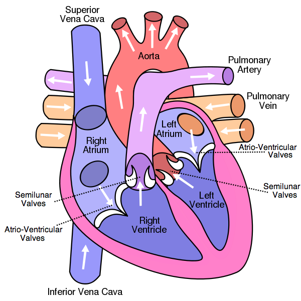
Figure 6.2.1 - The human heart
6.2.2 State that the coronary arteries supply heart muscle with oxygen and nutrients.
The coronary arteries supply heart muscle with oxygen and nutrients.
6.2.3 Explain the action of the heart in terms of collecting blood, pumping blood, and opening and closing of valves.
The right atrium collects blood from the superior and inferior vena cava and the left atrium collects blood from the pulmonary veins. This blood then flows into the right and left ventricle which pump the blood into the arteries. The direction of the blood flow is controlled by the atrioventricular valves and semilunar valves. When the atria contract the blood flows through the atrioventricular valves which are open, into the ventricle. At this stage the semilunar valves are closed so the ventricle fills with blood. The ventricles then contract which causes a rise in pressure. This rise in pressure first causes the atrioventricular valves to close preventing back flow of blood into the atria. Then the semilunar valves open allowing the expulsion of blood into the arteries. As this happens, the atria start to fill with blood again. The ventricles stop contracting leading to a fall in pressure which causes the semilunar valves to close, preventing back flow of blood from the arteries. When the ventricular pressure drops below the atrial pressure the atrioventricular valves open again and the cycle repeats.
Summary:
- Atria collect blood from veins.
- Atria contract, atrioventricular valves open.
- Blood is pumped into ventricles.
- Ventricle contracts, atrioventricular valves close and semilunar valves open.
- Blood is pumped into arteries, semilunar valves close.
- Cycle repeats.
6.2.4 Outline the control of the heartbeat in terms of myogenic muscle contraction, the role of the pacemaker, nerves, the medulla of the brain and epinephrine (adrenaline).
The heart muscle can contract by itself, without the stimulation of a nerve. This is called myogenic muscle contraction. The region that initiates each contraction is found in the wall of the right atrium and is called the pacemaker. Every time the pacemaker sends out a signal, a heartbeat results. The pacemaker is under the influence of nerves and adrenaline. One nerve carries messages from the medulla of the brain to the pacemaker and speeds up the beating of the heart. Another nerve carries messages from the medulla of the brain to the pacemaker and slows down the beating of the heart. Finally, adrenaline (epinephrine) is carried by the blood and once it reaches the pacemaker it signals it to increase the beating of the heart.
Summary:
- Heart muscle can contract by itself (myogenic muscle contraction).
- Pacemaker initiates contractions.
- One nerve carries messages from the brain to the pacemaker to speed up the beating of the heart.
- One nerve carries messages from the brain to the pacemaker to slow down the beating of the heart.
- Adrenaline signals the pacemaker to increase the beating of the heart.
6.2.5 Explain the relationship between the structure and function of arteries, capillaries and veins.
Arteries have a thick outer layer of longitudinal collagen and elastic fibers to avoid leaks and bulges. They have a thick wall which is essential to withstand the high pressures. They also have thick layers of circular elastic fibres and muscle fibres to help pump the blood through after each contraction of the heart. In addition the narrow lumen maintains the high pressure inside the arteries.
Veins are made up of thin layers with a few circular elastic fibres and muscle fibres. This is because blood does not flow in pulses and so the vein walls cannot help pump the blood on. Veins also have thin walls which allows the near by muscles to press against them so that they become flat. This helps the blood to be pushed forwards towards the heart. There is only a thin outer layer of longitudinal collagen and elastic fibres as there is low pressure inside the vein and so little chance of bursting. Finally, a wide lumen is needed to accommodate the slow flowing blood due to the low pressure.
Capillaries are made up of a wall that is only one cell layer thick and results in the distance for diffusion in and out of the capillary being very small so that diffusion can occur rapidly. They also contain pores within the their wall which allow some plasma to leak out and form tissue fluid. Phagocytes can also pass through these pores to help fight infections. In addition, the lumen of the capillaries is very narrow. This means that many capillaries can fit in a small space, increasing the surface area for diffusion.
Summary:
Arteries:
- Thick outer layer of longitudinal collagen and elastic fibres prevents leaks and bulges.
- Thick wall withstands high pressure.
- Thick layers of circular elastic fibres and muscle fibres to pump blood.
- Narrow lumen to maintain high pressure.
Veins:
- Thin layer with few circular elastic fibres and muscle fibres as blood does not flow in pulses.
- Thin walls so that nearby muscles can help push blood towards the heart.
- Thin outer layer of longitudinal collagen and elastic fibers as pressure is low.
- Wide lumen to accomodate the slow flowing blood.
Capillaries:
- Wall is one cell layer thick so distance for diffusion is small.
- Pores allow plasma to leak out and form tissue fluid. Phagocytes can also pass through pores.
- Very narrow lumen so that many can fit in a small space.
6.2.6 State that blood is composed of plasma, erythrocytes, leucocytes (phagocytes and lymphocytes) and platelets.
Blood is composed of plasma, erythrocytes, leucocytes (phagocytes and lymphocytes) and platelets.
6.2.7 State that the following are transported by the blood: nutrients, oxygen, carbon dioxide, hormones, antibodies, urea and heat.
Nutrients, oxygen, carbon dioxide, hormones, antibodies, urea and heat are all transported by the blood.