
Nerves, hormones and homeostasis
6.5.1 State that the nervous system consists of the central nervous system (CNS) and peripheral nerves, and is composed of cells called neurons that can carry rapid electrical impulses.
The nervous system consists of the central nervous system (CNS) and peripheral nerves, and is composed of cells called neurons which carry rapid electrical impulses.
6.5.2 Draw and label a diagram of the structure of a motor neuron.
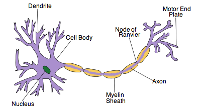
Figure 6.5.1 - A motor neuron
6.5.3 State that nerve impulses are conducted from receptors to the CNS by sensory neurons, within the CNS by relay neurons, and from the CNS to effectors by motor neurons.
Nerve impulses are conducted from receptors to the CNS by sensory neurons, within the CNS by relay neurons, and from the CNS to effectors by motor neurons.
6.5.4 Define resting potential and action potential (depolarization and repolarization).
Resting potential: the electrical potential across the plasma membrane of a cell that is not conducting an impulse.
Action potential: the reversal and restoration of the electrical potential across the plasma membrane of a cell, as an electrical impulse passes along it (depolarization and repolarization).
6.5.5 Explain how a nerve impulse passes along a non-myelinated neuron.
Sodium is found in greater concentrations outside of the cell while potassium is found in greater concentrations inside the cell. Sodium-potassium pumps exist in the plasma membrane to maintain the the concentration gradients and the membrane potential. Nerve impulses have a domino effect. An action potential in one part of the neuron causes another action potential in the adjacent part and so on. This is due to the diffusion of sodium ions between the region of the action potential and the resting potential. It is the movement of sodium and potassium that reduce the resting potential.
If the resting potential rises above the threshold level, voltage gated channels open. Voltage gated sodium channels open very fast so that sodium can diffuse into the cell down its concentration gradient. This reduces the membrane potential and results in more sodium channels opening. Sodium ions are positively charged and so the inside of the cell develops a net positive charge compared to the outside of the cell. This results in depolarization as the potential across the membrane is reversed.
A short while after this, voltage gated potassium channels open and potassium ions flow out of the cell down the concentration gradient. Since potassium ions are positively charged, their diffusion out of the cell causes a net negative charge to develop again inside the cell compared to the outside. The potential across the membrane is restored. This is called repolarization.
Finally, the concentration gradients of both ions are restored by the sodium-potassium pump. Sodium is pumped out of the cell while potassium is pumped in. The resting potential is restored and the neuron is ready to conduct another nerve impulse.
Summary:
- Resting potential rises above threshold level.
- Voltage gated sodium channels open.
- Sodium ions flow into the cell, more sodium channels open.
- Inside of cell develops a net positive charge compared to the outside and results in depolarization.
- Voltage gated potassium channels open.
- Potassium ions flow out of the cell.
- Cell develops a net negative charge compared to the outside and results in repolarization.
- Concentration gradients restored by sodium-potassium pumps.
- Resting potential is restored.
6.5.6 Explain the principles of synaptic transmission.
A synapse is a junction that permits a neuron to pass an electrical or chemical signal to another cell. At a synapse, the plasma membrane of the signal passing neuron (presynaptic neuron) is closely related to the plasma membrane of the target cell (postsynaptic neuron). Between the two there is a narrow fluid filled space called the synaptic cleft. Chemical signals called neurotransmitters pass from the presynaptic neuron to the post synaptic neuron.
This is how a synaptic transmission occurs:
An action potential travels along the neuron and reaches the end of the pre-synaptic neuron. The depolarization of the pre-synaptic membrane results in the opening of voltage gated calcium channels. Calcium ions flow into the presynaptic neuron and cause vesicles with neurotransmitters inside the neuron to fuse with the plasma membrane and release the neurotransmitters into the synaptic cleft via exocytosis. These neurotransmitters then diffuse within the synaptic cleft and some will bind to specific receptors located on the postsynaptic plasma membrane. The receptors are transmitted-gated ion channels which open and let sodium and other positively charged ions into the postsynaptic neuron when the neurotransmitters bind. As these positively charged ions enter the postsynaptic neuron they cause its membrane to depolarize. This depolarization results in an action potential which passes down the postsynaptic neuron. The neurotransmitters in the synaptic cleft are then quickly degraded and the calcium ions are pumped back into the synaptic cleft from inside the presynaptic neuron.
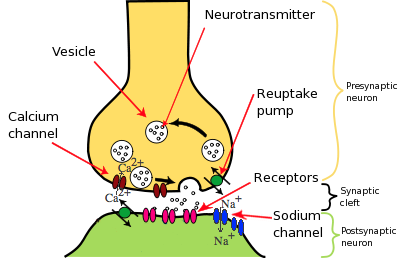
Figure 6.5.2 - Synaptic transmission
Summary:
- Action potential reaches the end of a presynaptic neuron.
- Voltage gated calcium channels open.
- Calcium ions flow into the presynaptic neuron.
- Vesicles with neurotransmitters inside the presynaptic neuron fuse with the plasma membrane.
- Neurotransmitters diffuse in the synaptic cleft and bind to receptors on the postsynaptic neuron.
- The receptors are channels which open and let sodium ions into the postsynaptic neuron.
- The sodium ions cause the postsynaptic membrane to depolarize.
- This causes an action potential which passes down the postsynaptic neuron.
- Neurotransmitters in the synaptic cleft are degraded and the calcium ions are pumped back into the synaptic cleft.
6.5.7 State that the endocrine system consists of glands that release hormones that are transported in the blood.
The endocrine system consists of glands that release hormones that are transported in the blood.
6.5.8 State that homeostasis involves maintaining the internal environment between limits, including blood pH, carbon dioxide concentration, blood glucose concentration, body temperature and water balance.
Homeostasis involves maintaining the internal environment between limits, including blood pH, carbon dioxide concentration, blood glucose concentration, body temperature and water balance.
6.5.9 Explain that homeostasis involves monitoring levels of variables and correcting changes in levels by negative feedback mechanisms.
Homeostasis involves maintaining the internal environment between limits, including blood pH, carbon dioxide concentration, blood glucose concentration, body temperature and water balance. Blood and tissue fluid (derived from blood) make up the internal environment. This internal environment varies very little compared to the external environment which varies greatly. Negative feed back is used to keep the internal environment between limits. It uses the nervous and endocrine system to do so. It has a stabilising effect as any change from a set point level will result in an opposite change. The levels of production of for example blood glucose, feed back to affect the rate of production. If blood glucose levels rise above the set point, this will feed back to decrease production and reduce the level back around the set point. A decrease in blood glucose levels below the set point will result in an increase in production so that the levels increase back to the set point. Small fluctuations around the set point will not cause any response. Negative feed back is only triggered when there are significant increases or decreases from the set point.
Summary:
- Homeostasis maintains the internal environment between limits.
- Negative feed back is used to do so. Any change from a set point results in an opposite change.
6.5.10 Explain the control of body temperature, including the transfer of heat in blood, and the roles of the hypothalamus, sweat glands, skin arterioles and shivering.
The hypothalamus is responsible for monitoring the temperature of the blood which is normally close to 37 degrees. If there are significant fluctuations from this set point, the hypothalamus sends signals (messages carried by neurons) to different parts of the body to restore the temperature back to the set point. This is done through negative feedback.
|
If blood temperature significantly increases above the set point |
If blood temperature significantly drops bellow the set point |
|
Skin arterioles increase in diameter so that more blood flows to the skin. By doing so it transfers heat from the core of the body to the skin and this heat is then lost to the external environment, cooling down the body in the process. |
Skin arterioles decrease in diameter so that less blood flows to the skin. The diameter of the capillaries in the skin cannot change but less blood flows through them. This prevents heat loss to the external environment as the temperature of the skin falls. |
|
Skeletal muscle stays relaxed so that more heat is not generated. |
Shivering occurs. This is when the skeletal muscle does many small rapid contractions to generate heat. |
|
Sweat glands secrete large amounts of sweat which makes the surface of the skin moist. When water evaporates from the moist skin it cools down the body. |
Sweat glands to not secrete sweat and so no water evaporation can occur as skin stays dry. |
6.5.11 Explain the control of blood glucose concentration, including the roles of glucagon, insulin and α and β cells in the pancreatic islets.
Blood glucose concentration does not have a specific set point like blood temperature. Blood glucose levels drop and rise through the day and so the body usually tries to keep blood glucose levels around 4 to 8 millimoles per dm3 of blood. Once again, negative feedback is used to do so. There are responses by target organs which affect the rate at which glucose is taken up from the blood or loaded into the blood.
|
Response to blood glucose levels above the set point |
Response to blood glucose levels below the set point |
|
β cells in the pancreatic islets produce insulin. Insulin stimulates muscle cells and the liver cells to take up glucose from the blood and convert it into glycogen. These are then stored in the form of granules in the cytoplasm of cells. Also, other types of cells are stimulated to take up glucose and use it for cell respiration instead of fat. All of these processes lower the levels of glucose in the blood. |
α cells in the pancreatic islets produce glucagon. Glucagon stimulates the liver cells to convert glycogen back into glucose and release this glucose into the blood. This raises the glucose levels in the blood. |
6.5.12 Distinguish between type I and type II diabetes.
|
Type I diabetes |
Type II diabetes |
|
The onset is usually early, sometime during childhood. |
The onset is usually late, sometime after childhood. |
|
β cells do not produce enough insulin. |
Target cells become insensitive to insulin. |
|
Diet by itself cannot be used to control the condition. Insulin injections are needed to control glucose levels. |
Insulin injections are not usually needed. Low carbohydrate diet can control the condition. |