Discuss the use of technology in investigating cognitive processes.
Introduction
- State what you are doing in the essay
- The following essay will attempt to offer a balanced review of the use of technology in investigating cognitive processes.
- State the different types of brain imaging technologies
- PET: Positron Emission Topography
- MRI: Magnetic Resonance Imaging
- fMRI: functional Magnetic Resonance Imaging EEG: Electroencephalogram
- CAT: Computerised Axial Tomography
- Each method has its own advantages and disadvantages and are appropriate in varying situations
- Explain why Brain imaging technologies are used at the CLA
- Brain imaging technologies are methods used in psychology to examine the human brain.
- Brain imaging technologies are quite useful in neuropsychology...
- As it provides an opportunity to study the active brain
- Allows researchers to see where specific brain processes take place
- Predominantly used to define brain differences in groups while they perform cognitive tasks
- Enables researchers to study localisation of function in a living human brain
- State the cognitive processes being discussed
- The cognitive processes being discussed in this essay are:
- Memory
- Language
- State the brain imaging technology being discussed
- Magnetic Resonance Imaging (MRI)
- Positron Emission Tomography (PET)
- Example Response
- In the following essay, the brain imaging technology that will be discussed are MRI and PET Scans and will be investigated in terms of its role in investigating the correlations/relationships between cognitive processes of memory and language.
Body
Cognitive Process 1: MEMORYBrain Imaging Technology 1: MRI Scans
- Introduce the cognitive process of memory
- The first brain imaging technology, MRI scans, will be firstly investigated with the cognitive process of memory.
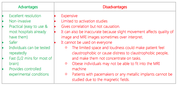
- Describe the MRI brain imaging technology
- This technique uses magnetic fields and radio waves to produce 3D computer-generated images.
- MRI scans involve people to remove all metal objects and clothing where they lie within an MRI machine.
- It can distinguish among different types of soft tissue and allows researchers to see structures within the brain.
Supporting Study: Maguire et al. (2000)
Introduce Study Connection of study to question:
- An example of a study which utilizes MRI scans to investigate the cognitive process of memory is a study conducted by Maguire et al. (2000).
- Maguire hypothesised that full licensed taxi drivers in London would have a different hippocampi structure in their brains compared to ‘normal’ people.
- This was based on the knowledge that London taxi drivers must do a two-year training course where they end up being able to find their way around the city without a map.
- MRI scans were used to scan the structure of their hippocampi, which were compared to already existing MRI scans of healthy males who did not drive taxis.
Results:
- Taxi drivers’ left and right hippocampi had a larger volume compared to the non-taxi drivers.
- Some parts of the hippocampi were smaller in the taxi drivers.
Conclusions:
- Maguire concluded that there was probably a redistribution of grey matter in the hippocampi of taxi drivers due to the regular use of the spatial memory skills required to remember roads; the neurons are stronger in areas of the brain which are used most.
- By using an MRI, Maguire was able to observe the structures in the brain and find a correlation between the hippocampi (biological factor) and memory skills (cognitive process).
- Maguire used MRI scans to investigate the structure of the hippocampi, which would not be able to be seen using other technologies such as an EEG or a PET scan
Supporting Study 2: HM Milner and Scoville (1957)
Introduce StudyConnection of study to question:
- Another study which utilizes MRI scans to investigate memory is a study conducted by Milner and Scoville (1957).
- Background:
- HM suffered epileptic seizures after a head injury at age 9
- Doctors performed surgery to stop seizures
- Tissue from temporal lobe, and hippocampus was removed
- HM suffered anterograde amnesia
- He could recall information from early life but could not form new memories
- HM was studied using an MRI in 1997
- Findings:
- The brain scan showed that there was damage to the hippocampus, amygdala, and areas close to the hippocampus
- By using MRI scanning technology, researchers were able to investigate the cognitive process of memory and make a correlation between certain brain areas (biological factor) and memory (cognitive process).
- MRI scans were used to see the structures of the brain to determine the extent of brain damage
- The structures would not be able to be clearly seen using other technologies such as EEGs or CTs.
Brain Imaging Technology 2: PET Scans
- Introduce the cognitive process of language
- The next cognitive process which will be discussed with the brain imaging technology of PET Scans is language.
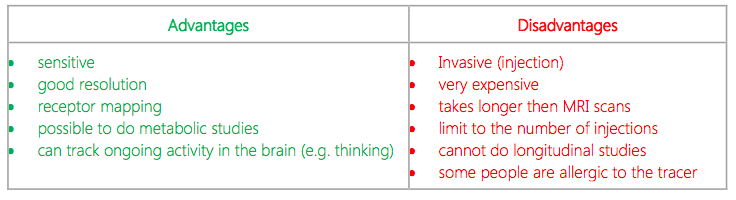
- Describe PET brain imaging technology
- PET scans require patients to be injected with a radioactive glucose tracer which shows the areas where glucose is absorbed in the active brain.
- More glucose metabolism means more brain activity.
- PET scans show a coloured visual display of brain activity; where radioactive tracer is absorbed
- Red indicates areas with the most activity
- Blue indicates areas with the least activity
Supporting Study 3: Tierney et al (2001)
Introduce Study --> Connection of study to question:
- An example of a study which utilizes PET scans to investigate the cognitive process of language is a study conducted by Tierney et al. (2001).
- To evaluate, using PET scans, the bilingual language compensation following early childhood brain damage
- 37 year old man (known as MA) with normal speech functions who was participating in a normal speech study
- It was discovered that he had a lesion in his left frontal lobe
- Probably as a result of encephalitis he suffered at the age of 6 weeks
- He had no significant long-term, clinically consequences
- Both his parents were deaf and he used sign language at home from a very young age.
- Researchers were curious to know if this might have had something to do with his ability to speak despite the brain damage (that should have prevented him from doing so.
- Researchers compared MA to 12 control participants, who were fluent in sign language
- PET scanning technologies were used while the participants produced narrative speech or signs
- MA's right hemisphere was more active than the controls' during the production of both speech and sign language
- Language function seems to have developed in the right hemisphere instead of the left hemisphere as an adaptation following his early brain damage
- Tierney utilised PET scans to investigate the cognitive processes of language and observe the areas of the brain (biological factor) that activated while MA produced language (cognitive process).
- The ongoing activity in the brain would not be able to be seen using other technologies such as EEGs or MRIs.
Conclusion
- What is the significance of using brain scans? Answer the question
- In conclusion, brain imaging technologies are very useful in investigating cognitive processes.
- Useful in different situations.
- All these methods have their own advantages and disadvantages, primarily involving invasiveness and levels of radioactivity.
- However, all of these methods contribute to investigating the relationship between cognitive processes and behaviour.
- It is important to note that different brain scans are used depending on the individual, the cause of the problem and or the cognitive process being investigated.